
Exams · Diagnostic imaging
Dermatologic ultrasound: high-frequency ultrasound of the skin
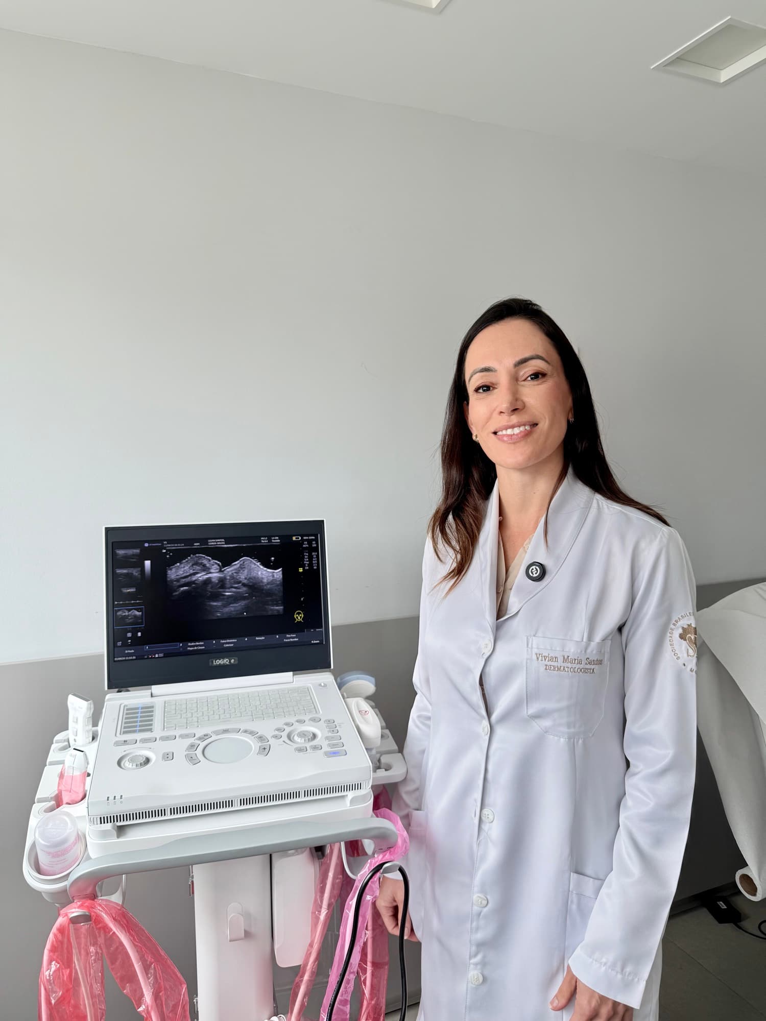
Dermatologic ultrasound is an imaging exam that uses high-frequency probes to show the layers of the skin, the nails, the scalp and the tissues just below, with no radiation and no pain. It helps diagnose lesions and tumors, monitor inflammatory diseases such as hidradenitis suppurativa, and locate products injected in aesthetic procedures on the face and buttocks. At Yamagata Dermatologia, in Ipanema, Rio de Janeiro, it is performed by a dermatologist.
- Duration
- 30 to 60 minutes
- Preparation
- Usually none
- Discomfort
- Painless and uses no radiation
- Result
- Report with images
How it works
The device emits ultrasound waves that pass through the skin and return as echoes, forming a real-time image of the structures below the surface. Dermatologic ultrasound uses high-frequency probes, which show the epidermis, dermis, the fat under the skin and the nails in detail.
Color Doppler, a feature of the same device, assesses the vascularization (blood flow) of lesions and surrounding tissues and identifies nearby blood vessels. This information helps distinguish types of nodules, assess the activity of inflammatory diseases and plan procedures.
Because it uses no radiation, the exam can be repeated whenever needed, which allows images to be compared throughout treatment.
Exams performed
- 01
Skin lesions and tumors
Shows the size, depth, margins and vascularization (blood flow) of skin nodules and tumors, which helps with diagnosis and planning surgical removal.
Indications
- Nodules and lumps in or just under the skin
- Cysts, lipomas and raised scars, such as keloids
- Skin tumors already diagnosed, to measure their extent before surgery
- 02
Nails
Assesses the nail plate, nail bed and matrix (the area under the skin where the nail is produced), which cannot be seen in the clinical exam. Regular nail polish does not need to be removed; gel nails and extensions should ideally be removed beforehand, because they hinder analysis of the nail plate, but they do not prevent assessment of the bed and matrix.
Indications
- Nodules, cysts and tumors under the nail
- Nail deformities and changes with no apparent cause
- Psoriasis of the nails and finger joints
- 03
Scalp
Assesses the thickness and structure of the scalp tissues, inflammatory changes, nodules and other lesions, as a complement to trichoscopy.
Indications
- Alopecia with inflammation or scarring
- Nodules and cysts on the scalp
- Monitoring of inflammatory scalp diseases
- 04
Hidradenitis suppurativa
Identifies nodules, fluid collections and tunnels (fistulas) under the skin, often larger than they appear in the clinical exam. It is used for diagnosis, staging (classification of severity) and monitoring of treatment.
Indications
- Confirming the diagnosis in early or doubtful cases
- Staging before defining treatment
- Monitoring the response to medical treatment, biologics or surgery
- 05
Facial mapping
For people who have had fillers or other facial procedures: locates the injected products, indicates the likely type of material and shows its relationship to blood vessels.
Indications
- Planning new procedures in people who already have fillers
- Nodules, swelling or inflammation after procedures
- Guiding hyaluronidase injection to dissolve hyaluronic acid
- Old fillers of unknown composition
- 06
Buttock mapping
For people who have had procedures on the buttocks: locates and measures the injected products and identifies nodules, fluid collections or inflammation.
Indications
- Planning new procedures in the buttock area
- Nodules, pain, hardening or asymmetry after procedures
- Products injected in the past of unknown composition
- 07
Soft tissues
Assesses deeper lesions, located in the fat under the skin and neighboring tissues.
Indications
- Lipomas and deep cysts
- Lumps under the skin of undetermined origin
- Fluid collections, inflammation and foreign bodies
What the exam is like
The patient lies down or sits, depending on the area examined. The doctor applies a gel to the skin and glides the probe over the area, recording images and measurements. At the end, the gel is removed and you can return to your normal activities.
The exam takes 30 to 60 minutes, depending on the number of areas assessed. The result is delivered in a report with images, which can be compared with future exams.
Preparation
Preparation is usually not needed. A few tips make the exam easier:
- Avoid creams, oils and makeup on the area to be examined on the day of the exam.
- Nails: regular nail polish does not need to be removed. Gel nails and extensions can hinder analysis of the nail plate and should ideally be removed before the exam, but they do not prevent assessment of the bed and matrix.
- Bring previous exams, such as ultrasound reports and biopsies.
- For facial and buttock mapping, provide the names of the products injected, the dates and the sites, if you know them.
Limitations
Ultrasound does not replace a biopsy in the diagnosis of tumors and has limited reach for very superficial, pigmented lesions, such as moles. In these cases, dermoscopy and total body mole mapping are the recommended exams, and the choice between them is made at the appointment.
All procedures are recommended and performed after an individual medical evaluation. Results vary from person to person.
Questions
Frequently asked questions
Is dermatologic ultrasound painful?
No. The exam is performed with a gel and a probe that glides over the skin, with no needles and no radiation. In inflamed areas, such as in hidradenitis suppurativa, there may be mild discomfort from the pressure of the probe.
What is the difference between dermatologic ultrasound and regular ultrasound?
Dermatologic ultrasound uses higher-frequency probes, which show structures close to the surface, such as the skin layers and the nail, in more detail. Conventional ultrasound reaches deeper organs, but shows less detail in these layers.
Does ultrasound replace a biopsy?
No. Ultrasound shows the size, depth and vascularization of a lesion and helps decide the approach, but the definitive diagnosis of a tumor depends on a biopsy and histopathological examination.
Can the exam identify which filler was injected?
In most cases, ultrasound distinguishes the main groups of materials, such as hyaluronic acid, biostimulators and permanent products, by their appearance on the image. Information about the product, date and site of injection makes the assessment more precise.
Can pregnant women and children have the exam?
Yes. Because it uses no radiation, dermatologic ultrasound can be performed during pregnancy and childhood, and repeated as often as needed for follow-up.
Do I need a doctor's referral?
Yes. Dermatologic ultrasound is performed with a medical request, which the dermatologist can issue at the appointment.
Who performs it
Physician in charge

Dr. Vivian Santos
Dermatologist
PHYSICIAN: CRM-RJ 52.88185-6 · Dermatologist: RQE 44331
Curriculum vitae
- Medical degree from the Federal University of the State of Rio de Janeiro (UNIRIO)
- Residency in Internal Medicine at the Hospital Municipal Miguel Couto
- Specialization in Dermatology at the Hospital Central do Exército (HCE)
- Postgraduate Diploma in Clinical Dermatology from Queen Mary University of London
- Residency in Leprology (Hansen's disease) at the Gaffrée e Guinle University Hospital (HUGG)
- Fellowship in Dermatologic Ultrasound at CETRUS
- Professor at the Instituto de Dermatologia IMS