
Exams · Diagnostic imaging
Dermoscopy: magnified examination of moles and skin lesions
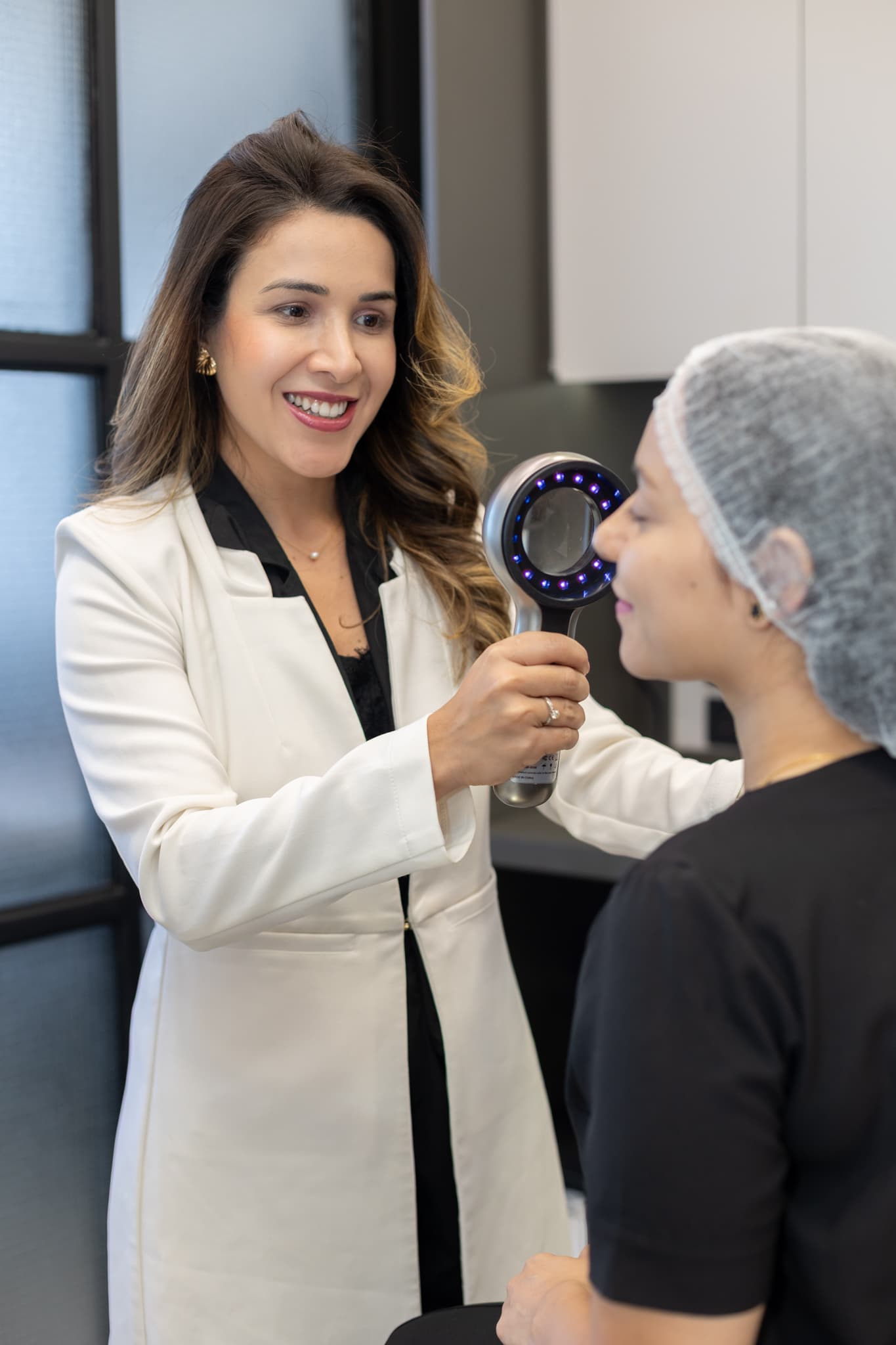
Dermoscopy is an in-office exam performed with a dermatoscope, a device that magnifies the image of the skin and uses special light to show structures and colors that are not visible to the naked eye. It helps distinguish benign moles and lesions from those suspected of skin cancer and is also used for diseases of the scalp, nails and other skin conditions. At Yamagata Dermatologia, in Ipanema, Rio de Janeiro, it is performed by dermatologists.
- Duration
- Performed during the appointment
- Discomfort
- Painless and uses no radiation
- Device
- Handheld or digital dermatoscope
- Preparation
- None required
How it works
The dermatoscope combines a magnifying lens with special light, polarized or not, that reduces reflection from the skin surface. This lets the doctor see the distribution of pigment, blood vessels and other structures below the surface, which form patterns characteristic of each type of lesion.
Dermoscopy can be performed with a handheld device, during the appointment, or with a digital dermatoscope, which photographs and stores the images for comparison, as in total body mole mapping.
What it is used for
- Distinguishing benign moles from lesions suspected of melanoma.
- Recognizing other skin cancers, such as basal cell carcinoma, and benign lesions, such as seborrheic keratosis.
- Assessing the scalp and hair (trichoscopy) and the nails (onychoscopy).
- Aiding the diagnosis of inflammatory and infectious skin diseases.
What the exam is like
The doctor places the dermatoscope on the lesion, sometimes with a drop of gel or alcohol, and analyzes the magnified image. The exam is performed during the appointment, is painless and requires no preparation.
Results
The result is discussed at the appointment itself: the lesion may be considered benign, monitored with new images or recommended for removal and analysis, through dermatologic surgery.
Limitations
Dermoscopy increases the accuracy of the clinical exam, but does not replace a biopsy when there is doubt: the definitive diagnosis of a suspicious lesion is made by histopathological examination.
All procedures are recommended and performed after an individual medical evaluation. Results vary from person to person.
Questions
Frequently asked questions
What is the difference between dermoscopy and total body mole mapping?
Dermoscopy is the examination of lesions with a dermatoscope, performed during the appointment. Total body mole mapping is a more complete exam that photographs the entire skin and stores dermoscopic images of the moles for comparison over time.
Does dermoscopy avoid biopsies?
It helps avoid unnecessary biopsies, because it allows benign lesions to be recognized with more confidence, and helps indicate the removal of suspicious lesions more precisely.
Is dermoscopy used only for moles?
No. It is also used to assess other skin lesions, the scalp (trichoscopy), the nails (onychoscopy) and some inflammatory and infectious skin diseases.
Team