
Exams · Diagnostic imaging
Digital total body mole mapping: mole monitoring with FotoFinder®
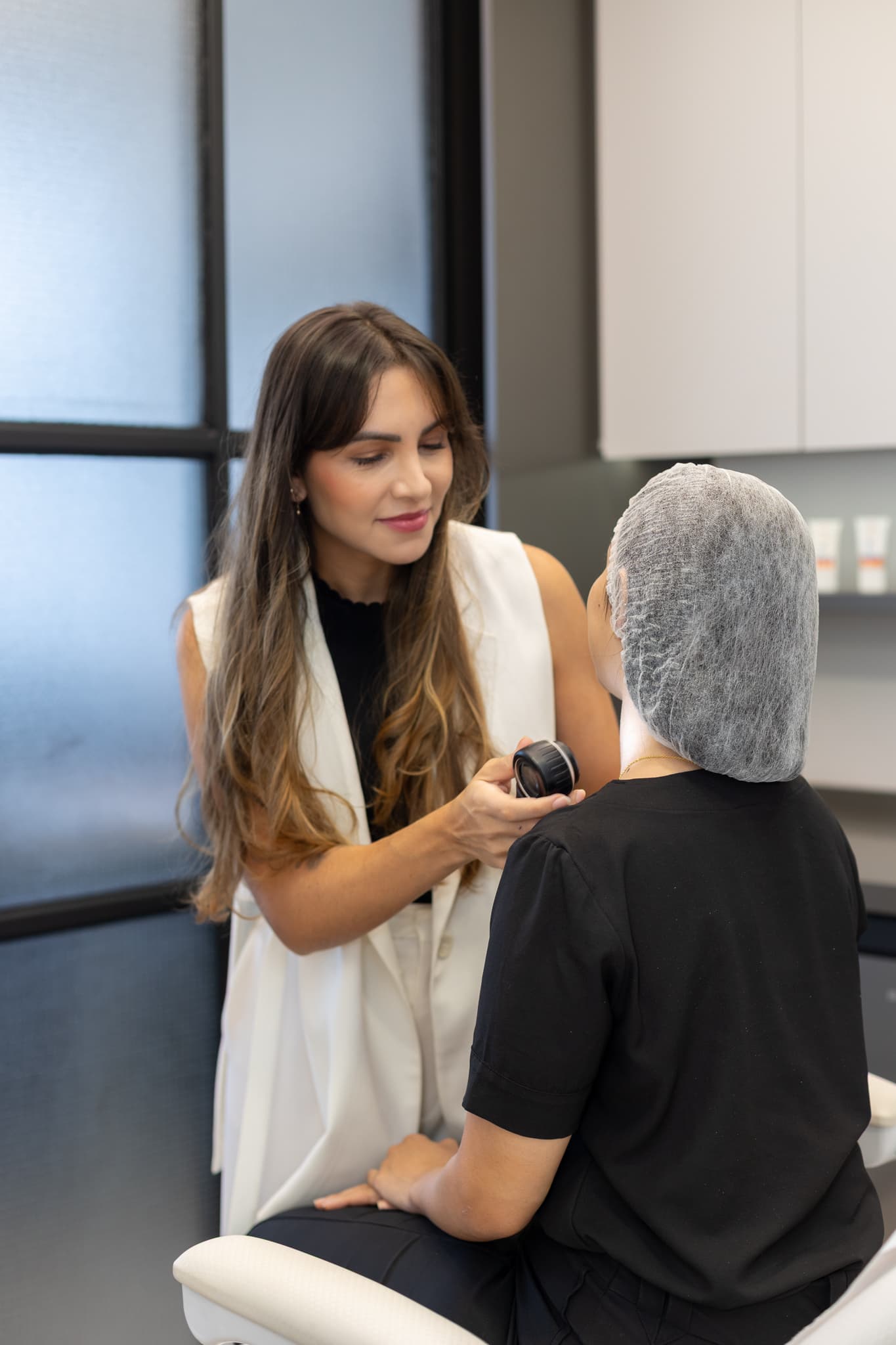
Digital total body mole mapping is an exam that photographs the entire skin surface in standardized positions and records moles with digital dermoscopy, which magnifies each lesion. At the clinic, it is performed with the FotoFinder® system. By comparing the images from one exam with those of the next, the dermatologist identifies new or changing moles, which allows early diagnosis of melanoma and other skin cancers. At Yamagata Dermatologia, in Ipanema, Rio de Janeiro, it is performed by dermatologists.
- Duration
- About 1 hour
- System
- FotoFinder®
- Discomfort
- Painless and uses no radiation
- Result
- Report and images for comparison
How it works
The exam has two stages, performed with the FotoFinder® system:
- Total body photography: the body is photographed in standardized positions, like a map of the skin. In later exams, the system compares the images and helps identify moles that have appeared or changed in size.
- Digital dermoscopy: the moles selected by the doctor are photographed with a lens that magnifies them up to 140 times, showing structures that are not visible to the naked eye. The images are stored and compared over time.
The system includes automated image comparison features that assist the doctor, but the interpretation and the decision about each lesion are always the dermatologist's.
Who it is recommended for
Total body mole mapping is recommended for people at higher risk of skin cancer or whose moles are difficult to monitor with the clinical exam alone:
- Many moles (generally more than 50) or atypical moles.
- Personal or family history of melanoma.
- Very fair skin, freckles, light eyes and a history of sunburn.
- Use of medications that lower immunity, such as after transplants.
What the exam is like
The patient wears underwear, in an area prepared for the photographs, and takes the indicated positions. The doctor then examines the skin and records with digital dermoscopy the moles that need monitoring. The exam takes about 1 hour.
Preparation
- Come without makeup, without creams or oils on your body and without dark nail polish.
- Avoid tanning and self-tanner in the preceding weeks.
- Bring previous exams and reports, if any.
Results and follow-up
The result includes the recorded images and guidance on each lesion: monitoring, re-evaluation at a short interval or removal for analysis. The date of the next exam is set according to each person's risk.
Limitations
Mole mapping does not replace the clinical exam or a biopsy: the definitive diagnosis of a suspicious lesion is made by histopathological examination, after removal through dermatologic surgery. Areas such as the scalp and mucous membranes are assessed in the clinical exam.
All procedures are recommended and performed after an individual medical evaluation. Results vary from person to person.
Questions
Frequently asked questions
Who should have total body mole mapping?
The exam is recommended for people with many moles (generally more than 50), atypical moles, a personal or family history of melanoma, very fair skin with freckles and a history of sunburn, or who take medications that lower immunity. The indication is made at the appointment.
How often should I repeat the exam?
The frequency depends on each person's risk and is defined by the dermatologist, ranging from a few months, to monitor a specific lesion, to a yearly interval.
Does mole mapping replace the appointment with the dermatologist?
No. Mole mapping is a documentation and comparison tool. The interpretation of the images and the clinical skin exam are carried out by the dermatologist.
Is the exam painful or does it require preparation?
It is painless and uses no radiation. Come without makeup, without dark nail polish and with no creams on your skin, and avoid tanning before the exam.
What happens if a suspicious mole is found?
The mole may be monitored with new images at a short interval or removed for analysis, depending on the evaluation. Removal is done through dermatologic surgery, with histopathological examination of the lesion.
Team